Autopsies Show COVID-19 Virus in Brain and Elsewhere in Body - Neuroscience News | By The Perfect Enemy
Summary: Autopsy tissue samples of 44 people who died of COVID-19 showed SAR-CoV-2, the virus responsible for coronavirus, spread throughout the body and to the brain, with traces of the virus lingering for 8 months.
Source: University of Minnesota
An analysis of tissue samples from the autopsies of 44 people who died with COVID-19 shows that SAR-CoV-2 virus spread throughout the body—including into the brain—and that it lingered for almost eight months.
The study was published in Nature.
Scientists from the National Institutes of Health (NIH) tested samples from autopsies that were performed from April 2020 to March 2021. They conducted extensive sampling of the nervous system, including the brain, in 11 of the patients.
RNA and viable virus in various organs
All of the patients died with COVID-19, and none were vaccinated. The blood plasma of 38 patients tested positive for SARS-CoV-2, three tested negative, and plasma was unavailable for the other 3.
Thirty percent of the patients were female, and the median age was 62.5 years. Twenty-seven patient (61.4%) had three or more comorbidities. The median interval from symptom onset to death was 18.5 days.
Analysis showed that SARS-CoV-2, as expected, primarily infected and damaged airway and lung tissue. But the researchers also found viral RNA in 84 distinct body locations and bodily fluids, and in one case they isolated viral RNA 230 days after a patient’s symptoms began.
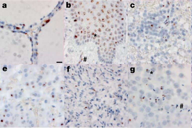
The researchers detected SARS-CoV-2 RNA and protein in the hypothalamus and cerebellum of one patient and in the spinal cord and basal ganglia of two other patients. But they found little damage to brain tissue, “despite substantial viral burden.”
The investigators also isolated viable SARS-CoV-2 virus from diverse tissues in and outside the respiratory tract, including the brain, heart, lymph nodes, gastrointestinal tract, adrenal gland, and eye. They isolated virus from 25 of 55 specimens tested (45%).
The authors wrote, “We demonstrated virus replication in multiple non-respiratory sites during the first two weeks following symptom onset.”

They add, “Our focus on short postmortem intervals, a comprehensive standardized approach to tissue collection, dissecting the brain before fixation, preserving tissue in RNA later, and flash freezing of fresh tissue allowed us to detect and quantify SARS-CoV-2 RNA levels with high sensitivity by [polymerase chain reaction] and [in situ hybridization], as well as isolate virus in cell culture from multiple non-respiratory tissues including the brain, which are notable differences compared to other studies.”
Possible ramifications for long COVID
Senior study author Daniel Chertow, MD, MPH, said in an NIH news release that, prior to the work, “the thinking in the field was that SARS-CoV-2 was predominantly a respiratory virus.”
Finding viral presence throughout the body—and sharing those findings with colleagues a year ago—helped scientists explore a relationship between widely infected bodily tissues and “long COVID,” or symptoms that persist for weeks and months after infection.
Part of a Paxlovid RECOVER trial that is expected to begin in 2023 includes an extension of the autopsy work highlighted in the Nature study, according to coauthor Stephen Hewitt, MD, Ph.D., who serves on a steering committee for the RECOVER project. Autopsies in the RECOVER trial include people who both were vaccinated and infected with variants of concern—data that weren’t available in yesterday’s study.
“We’re hoping to replicate the data on viral persistence and study the relationship with long COVID,” Hewitt said. “Less than a year in, we have about 85 cases, and we are working to expand these efforts.”
About this COVID-19 and neurology research news
Author: Jim Wappes
Source: University of Minnesota
Contact: Jim Wappes – University of Minnesota
Image: The image is credited to the researchers
Original Research: Closed access.
“SARS-CoV-2 infection and persistence in the human body and brain at autopsy” by Sydney R. Stein et al. Nature
Abstract
SARS-CoV-2 infection and persistence in the human body and brain at autopsy
Coronavirus disease 2019 (COVID-19) is known to cause multi-organ dysfunction during acute infection with severe acute respiratory syndrome coronavirus 2 (SARS-CoV-2), with some patients experiencing prolonged symptoms, termed post-acute sequelae of SARS-CoV-2.
However, the burden of infection outside the respiratory tract and time to viral clearance are not well characterized, particularly in the brain.
Here we carried out complete autopsies on 44 patients who died with COVID-19, with extensive sampling of the central nervous system in 11 of these patients, to map and quantify the distribution, replication and cell-type specificity of SARS-CoV-2 across the human body, including the brain, from acute infection to more than seven months following symptom onset.
We show that SARS-CoV-2 is widely distributed, predominantly among patients who died with severe COVID-19, and that virus replication is present in multiple respiratory and non-respiratory tissues, including the brain, early in infection. Further, we detected persistent SARS-CoV-2 RNA in multiple anatomic sites, including throughout the brain, as late as 230 days following symptom onset in one case.
Despite extensive distribution of SARS-CoV-2 RNA throughout the body, we observed little evidence of inflammation or direct viral cytopathology outside the respiratory tract. Our data indicate that in some patients SARS-CoV-2 can cause systemic infection and persist in the body for months.
#Coronavirus, #Coverage
Published on The Perfect Enemy at https://bit.ly/3QlJNUR.

Comments
Post a Comment
Comments are moderated.